


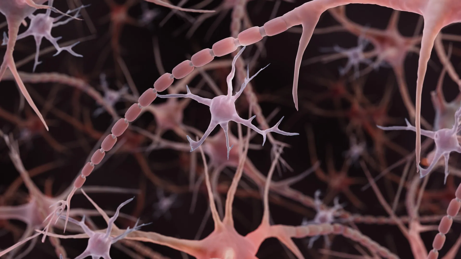
The brain’s microscopic wiring may need a different kind of textbook illustration. Instead of the smooth, narrow tubes commonly drawn extending from nerve cells, some axons resemble strings of tiny pearls. These repeating bulges are not simply decorative details. Their shape appears to influence how electrical messages travel.
Researchers at Johns Hopkins Medicine brought this unexpected architecture into focus in a study published online in Nature Neuroscience on December 2, 2024. Working with mouse neurons, they found evidence that a familiar picture of brain cell anatomy may overlook an important feature of the cells’ signal-carrying extensions.
Related research has since documented pearled axons in human brain tissue, extending the observations beyond mice. Together, the findings raise questions about how the physical structure of the brain’s wiring helps regulate communication.
“Understanding the structure of axons is important for understanding brain cell signaling,” said Shigeki Watanabe, Ph.D., an associate professor of cell biology and neuroscience at the Johns Hopkins University School of Medicine, in the university’s 2024 announcement. “Axons are the cables that connect our brain tissue, enabling learning, memory and other functions.”
A Different Picture of Brain Cell Axons
Axons are the long extensions that allow neurons to transmit electrical signals to other cells. The conventional illustration shows a relatively uniform tube interrupted by occasional bulges (synaptic varicosities that hold globs of neurotransmitters, which enable signaling to other brain cells).
Beaded axons themselves are not an unfamiliar sight. Scientists have long observed pronounced swellings in dying neurons and in neurodegenerative conditions, including Parkinson’s disease. In those settings, the beading can accompany damage to the cell membrane and the internal framework that helps maintain the axon’s structure.
What stood out in the Johns Hopkins study was the presence of much smaller, repeating swellings in axons examined under conditions intended to preserve their normal structure. The researchers called these regions “non-synaptic varicosities.” Unlike the familiar bulges associated with communication sites, these swellings were not synapses.
The distinction matters: the study did not suggest that every beaded axon is damaged. Instead, it pointed to a form of nanoscale pearling that may be part of the normal architecture of the axons studied.
A Clue From Worms
Watanabe’s interest began with observations of repeating pearls along axons in worms. A conversation with Swiss scientist Graham Knott, Ph.D., prompted him to investigate further. One possible explanation involved the axon’s internal skeleton, a supporting network of proteins.
A Harvard University team had reported repeating “skeletal” components within axons in a study published in 2012. Watanabe and Knott wondered whether disrupting that framework would make the pearls disappear. Jacqueline Griswold, a Johns Hopkins graduate student and the study’s first author, tested the idea. The pearling remained.
That result redirected their attention toward the axon’s physical properties. Watanabe and Griswold joined forces with theoretical biophysicist Padmini Rangamani, Ph.D., a professor of pharmacology at the University of California San Diego School of Medicine, to investigate how the surrounding membrane might shape the axon.
Freezing Brain Cells Reveals Hidden Detail
Studying these extensions of brain cells (neurons) requires imaging structures far narrower than a human hair. Johns Hopkins described them as roughly 100 times smaller than a hair’s width. The researchers used high pressure freezing electron microscopy to preserve their delicate shapes before examining them with beams of electrons.
The preparation method was important. Standard electron microscopy commonly involves chemically fixing and dehydrating tissue, steps that can alter the structures scientists are trying to observe. Freezing offers a way to retain a shape closer to the original.
“To see nanoscale structures with standard electron microscopy, we fix and dehydrate the tissues, but freezing them retains their shape — similar to freezing a grape rather than dehydrating it into a raisin,” Watanabe said.
The team examined mouse neurons grown in the laboratory, neurons from adult mice and neurons from mouse embryos. Their axons were nonmyelinated (they were without the myelin insulating cover that surrounds the axon). Across tens of thousands of tissue images, the researchers repeatedly encountered the same pearled appearance.
They also observed pearling with high resolution imaging of living neurons, providing evidence that it was not simply produced by freezing the samples.
“These findings challenge a century of understanding about axon structure,” Watanabe said.
How Membrane Physics Shapes Axons
To explain the pattern, the researchers built mathematical models of the membrane surrounding an axon. Relatively simple mechanical models reproduced important features of the pearls, suggesting that physical forces could help determine the shape without requiring a rigid internal mold.
Experiments supported that interpretation. Increasing the sugar concentration in the solution surrounding the axons made the swollen regions smaller. In the model, increasing membrane tension also reduced their size.
Cholesterol provided another way to change the membrane’s behavior. Removing it made the membrane less stiff and more fluid. This altered the pearled structure in both the models and mouse neurons and reduced the speed of electrical signaling.
“A wider space in the axons allows ions [chemical particles] to pass through more quickly and avoid traffic jams,” Watanabe said.
However, bigger pearls did not invariably mean faster signals. The dimensions of both the swellings and their narrow connecting segments mattered.
Electrical Activity Changes the Pearls
The researchers also tested whether neural activity could reshape the axons. After high frequency electrical stimulation, the pearl-like regions became an average of 8% longer and 17% wider, with enlargement lasting at least 30 minutes.
The published paper reports that electrical signals slowed after this stimulation, with the effect lasting at least an hour. When cholesterol had been removed beforehand, some structural changes still occurred, but the pearls remained smaller than those in untreated cells, and the same significant slowing was not detected.
The findings suggest that axons are not merely fixed cables. Their physical structure can respond to activity and help adjust how messages move through them.
Later Research Finds Pearled Axons in Human Tissue
When the original findings were announced in 2024, the team planned to examine axonal “arms” in human brain tissue obtained with permission from people undergoing brain surgery and from people who had died with neurodegenerative diseases.
A related study subsequently reported pearled axons in human cortical tissue obtained during epilepsy surgery. Led by Chelsy R. Eddings and colleagues, including Watanabe, the work appeared online in Neuron on November 24, 2025, and in its February 4, 2026, issue.
The researchers combined electrical stimulation with rapid freezing to capture tiny membrane changes in mouse and human brain slices. Alongside the pearled axons, they found evidence of ultrafast endocytosis, a process through which nerve endings quickly retrieve membrane after releasing chemical messages.
These observations extend the evidence to human tissue, but they do not establish that every axon has the same shape. Nor do they show that pearling has identical effects on signaling throughout the human brain. The human study primarily investigated membrane recycling at synapses.
The Broader Search for What Controls Axon Shape
The relationship between axon structure and function also became the focus of a National Institute of Mental Health Multiple Principal Investigator grant awarded to Watanabe and Rangamani and highlighted in 2024. The project supports computational models of how a neuron’s physical properties and incoming signals affect its axon, followed by tests in mouse tissue and cell cultures.
Separate research is exploring other ways to investigate the dimensions of this microscopic wiring. In a study published in PLOS Biology on July 17, 2026, University of Edinburgh researchers used automated imaging of living zebrafish to screen 880 compounds for effects on axon diameter. They identified 33 initial hits and confirmed that compounds affecting particular chemical signaling pathways could increase axon width. This was not a direct replication of the pearling study, but it provides another tool for investigating how axon size is controlled.
For the Johns Hopkins team, an important question remains how the structure seen in functioning axons relates to the damaging beading associated with neurological disease. Understanding that distinction could help researchers separate normal changes in neural wiring from signs that the system is breaking down.















Leave a comment