


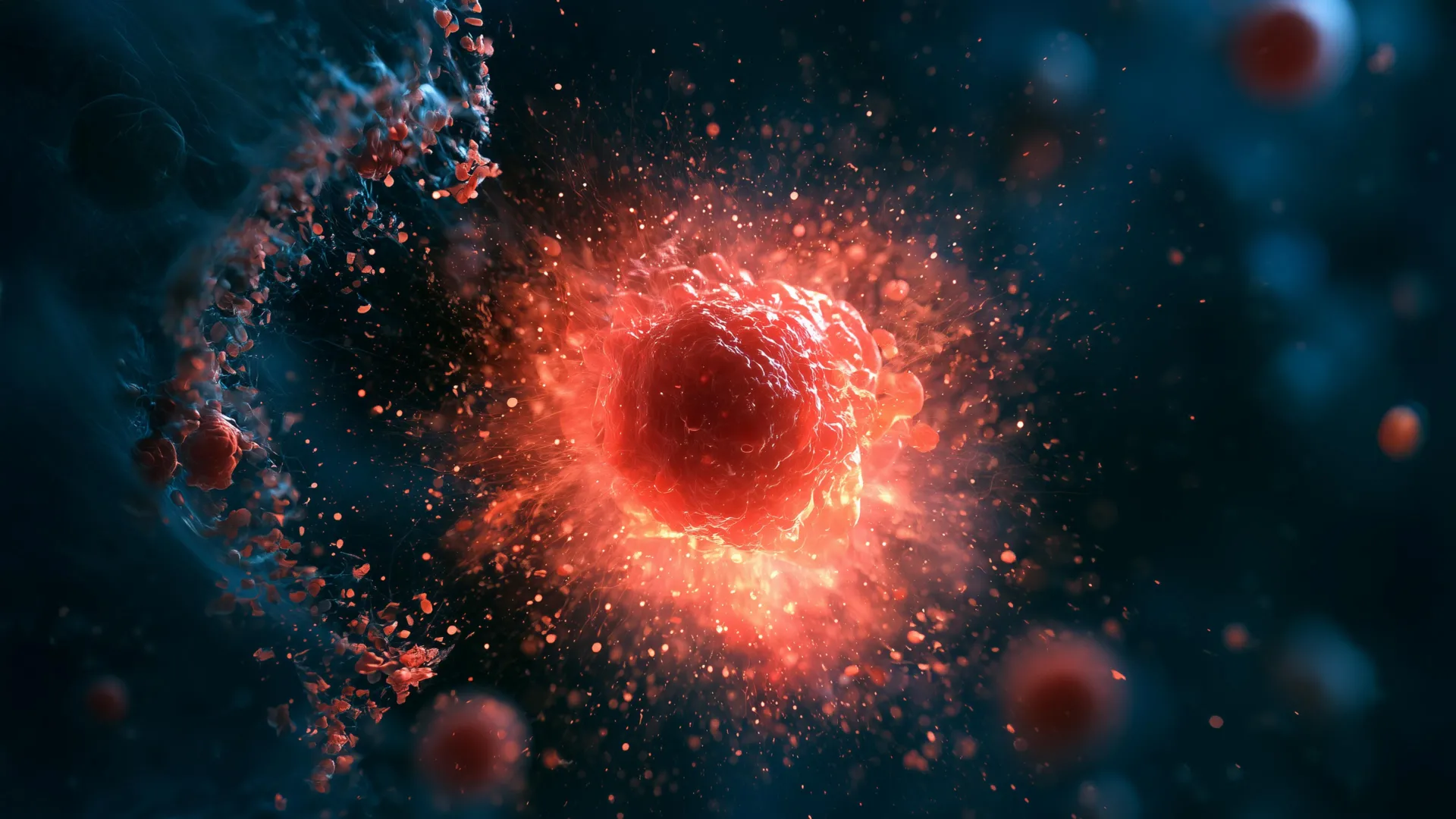
Stanford researchers have identified a previously unknown immune cell that destroys nearby cells by exploding. The process unfolds so rapidly and completely that the cell disappears within minutes, leaving virtually no trace.
The discovery was made in planarian flatworms, small aquatic animals known for their extraordinary ability to regenerate. Even when cut into pieces, these worms can rebuild missing tissues and, in some cases, produce complete new organisms. Studying how their immune systems have survived and adapted over hundreds of millions of years could offer useful clues for modern medicine.
In a study published in Cell, the researchers named the newly discovered cells “ruptoblasts” because of the dramatic way they respond to a specific hormone.
“We never expected that a cell could just explode like a bomb and kill the cells surrounding it,” said senior author Bo Wang, associate professor of bioengineering in the schools of Engineering and Medicine.
Flatworms Reject Foreign Tissue
Chew Chai, a postdoctoral researcher in Wang’s laboratory, first noticed the unusual cells while investigating a long-standing question in flatworm biology. Scientists wanted to know whether flatworms can distinguish their own tissues from those belonging to another individual.
To test this, Chai sliced worms lengthwise and fused each one with part of a different worm. Flatworms are highly effective at rebuilding their own bodies, but these fused “Frankenstein” worms rejected tissue from unrelated individuals. The reaction resembled the way a human immune system may reject a transplanted organ.
The cellular response, however, was very different from anything normally seen in humans.
“It’s this huge inflammatory response. Like there’s a fire and an alarm goes off, and the cells just blow up,” said Chai, who is lead author of the paper.
Activin Triggers Severe Inflammation
Previous research into flatworm regeneration has shown that the hormone activin plays an important role in their survival. Elevated activin levels can weaken a worm’s ability to regenerate, while low levels interfere with its ability to reproduce with other worms.
As the fused worms began rejecting foreign tissue, Chai detected a rise in activin followed by chronic inflammation. The animals did not die immediately, but they perished within several days. She also found that injecting activin into healthy, nonfused flatworms produced a similar inflammatory response.
To examine what was happening inside individual cells, Chai used live cell microscopy and flow cytometry, a technique that uses lasers to analyze and separate cells. She marked the cells with different fluorescent dyes and then isolated those that reacted to activin.
A small group of cells suddenly burst open, released substances that killed nearby cells, and disappeared within five minutes. Chai and Wang named this explosive process “ruptosis.”
Cell Death in Seconds
The speed and completeness of ruptoblast self-destruction distinguish ruptosis from other known forms of cell death.
“Some mammalian cells and bacteria may also do an explosive sort of cell death, but the timescale is really long. They are exploding, but it’s more like pores that slowly leak things out over the course of several hours,” said Chai. “Ruptosis happens within seconds to minutes.”
This rapid destruction appears to turn each ruptoblast into a highly localized weapon. Instead of slowly releasing harmful material, the cell unloads its contents almost instantly.
Explosive Cells Destroy Multiple Targets
Researchers tested ruptoblasts against E. coli bacteria, human kidney cells, and mouse blood cells. The ruptoblasts destroyed all three types of targets.
The damage remained limited to cells located close to the explosion. It did not spread through a chain reaction or leave behind persistent toxicity. According to Wang, this ability to deliver a powerful but tightly contained attack could have implications for future treatments targeting bacterial infections or tumors.
Ruptoblasts also differ from familiar immune cells such as T cells and neutrophils. Those cells are hematopoietic cells, meaning they are blood cells produced in bone marrow. Ruptoblasts are glandular cells.
The researchers believe ruptoblasts intensify their normal secretion systems so they can release toxic substances suddenly and violently after encountering activin. A rapid surge of calcium from the endoplasmic reticulum inside the cell helps drive ruptosis.
An Ancient Immune Strategy
When Chai searched for similar cells in other species, she found them only in basal bilaterians such as flatworms. Their restricted distribution suggests that ruptoblasts originated early in animal evolution.
Chai proposed that vertebrates may have lost this defense because they cannot easily repair the surrounding damage caused by ruptosis. Flatworms, by contrast, contain abundant stem cells and can replace damaged tissues with remarkable efficiency.
“It demonstrates there’s lots of different immune mechanisms out there. There’s all these animals that live in an environment where there’s lots of bacteria, lots of viruses, and we know so little about their immune mechanisms,” said Wang.
The findings show how much scientists may learn by examining animals that are rarely used as traditional research models. Although flatworms may appear simple, their unusual biology could reveal immune strategies that are absent from humans and other vertebrates.
Wang said that investigating a wider range of organisms may inspire new approaches to some of medicine’s most challenging problems.
Acknowledgements
Additional Stanford co-authors include postdoctoral scholar Souradeep Sarkar; former Undergraduate Visiting Research Program scholar Lihan Zhong; Dania Nanes Sarfati, PhD ’24; Christine Jacobs-Wagner, the Dennis Cunningham Professor and professor of biology in the School of Humanities and Sciences and of microbiology and immunology in the School of Medicine; and Hawa Racine Thiam, assistant professor of bioengineering in the schools of Engineering and Medicine and of microbiology and immunology in the School of Medicine. Additional co-authors, including co-senior author Benyamin Rosental, are from Ben Gurion University of the Negev.
Jacobs-Wagner is also a member of Stanford Bio-X and an institute scholar at Sarafan ChEM-H. Thiam is also a member of Bio-X and the Maternal & Child Health Research Institute (MCHRI), and an institute scholar at Sarafan ChEM-H. Wang is also a member of Bio-X and the Wu Tsai Neurosciences Institute.
This research was funded by a National Science Foundation Graduate Research Fellowship, a Stanford Graduate Fellowship, a Stanford DARE fellowship, a Human Frontier Science Program grant, a National Institutes of Health grant, and the European Research Council.














Leave a comment